

Multidimensional Molecular Imaging
We develop innovative instruments and technologies for multimodal bioimaging at the molecular level.
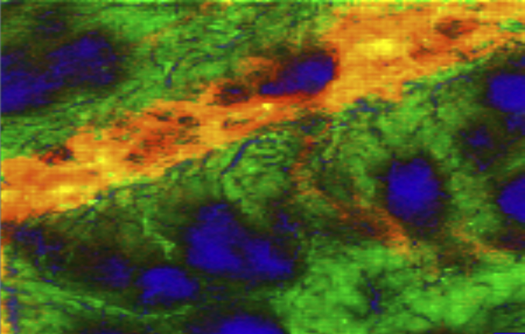
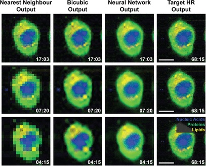
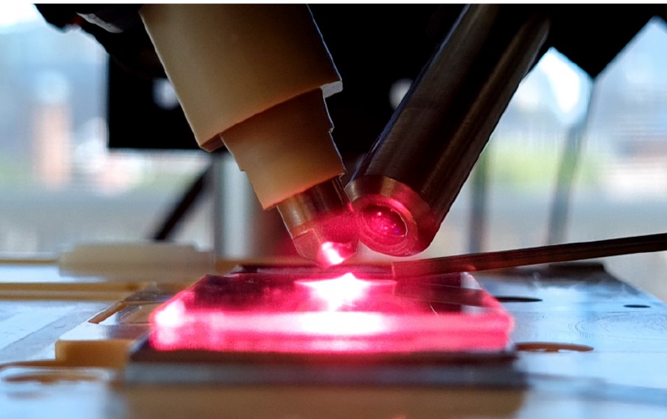
Label-free bioimaging harnesses direct interactions between excitation light and biological samples, including scattering, absorption, and fluorescence decay, to reveal biology without staining or labelling.
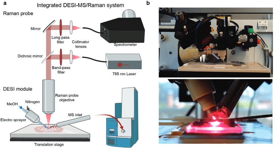
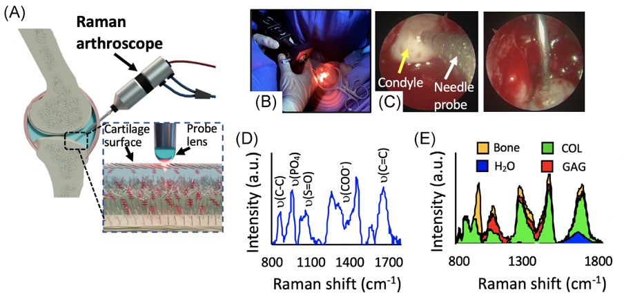
Our laboratory develops advanced correlative imaging approaches that combine multi-photon microscopy, fluorescence lifetime imaging (FLIM), confocal Raman spectroscopic imaging, and optical coherence tomography. We further integrate optical spectroscopy with DESI-mass spectrometry imaging and spatial transcriptomics for a comprehensive molecular view of biological systems.
Our laboratory develops advanced correlative imaging approaches that combine multi-photon microscopy, fluorescence lifetime imaging (FLIM), confocal Raman spectroscopic imaging, and optical coherence tomography. We further integrate optical spectroscopy with DESI-mass spectrometry imaging and spatial transcriptomics for a comprehensive molecular view of biological systems.